Experiments
Atom and molecular physics

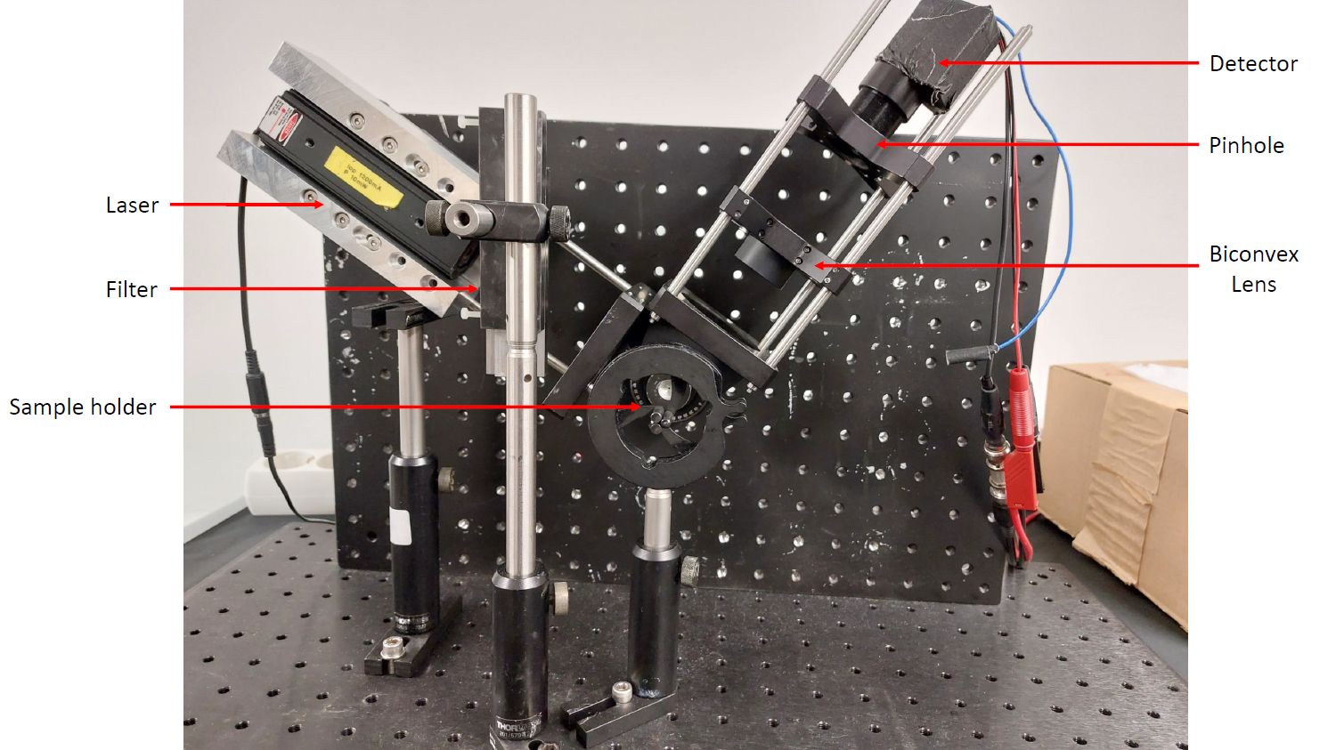
In the experiment, a helium-neon laser is set up and adjusted. Next, the power, polarization, spectral modes and spatial modes of the laser are investigated for different mirror arrangements and resonator lengths.
The experiment is suitable for all courses of study. Supervision can be provided in German or English.
Marlon Schäfer
Building E 2.6, Room 2.05
Phone: +49 681 302-3997
marlon.schaefer(at)physik.uni-saarland.de
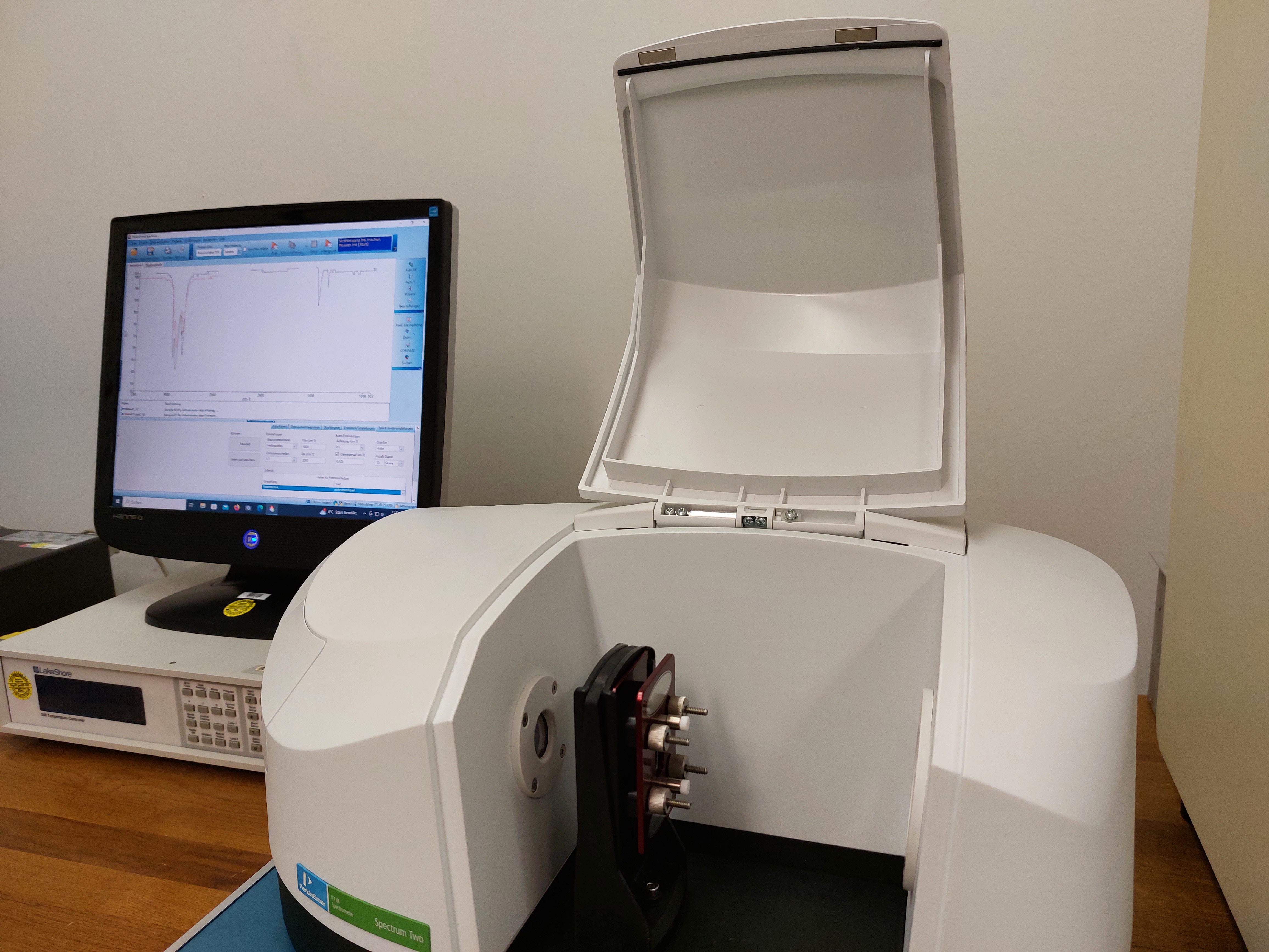
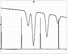
In the infrared spectroscopy experiment, you will become acquainted with the setup and functionality of an FTIR spectrometer as well as its various applications, and you will also learn how to prepare and handle samples in the experiment. In five experiments, you will determine the rotational and distortion constants of CO gas for different states, characterize materials, calculate refractive indices, and investigate phase transitions.
The experiment is suitable for all courses of study. Supervision can be provided in German or English.
Klaus Schappert
Building E 2.6, Room 2.20
Phone: +49 681 302-3032
k.schappert(at)mx.uni-saarland.de
In 1896 Peter Zeeman discovered the splitting of spectral lines of sodium when the light source was placed in a strong magnetic field, this phenomenon is called Zeeman effect. The Zeeman Effect was very important in the development of quantum mechanics, it provided direct evidence for the quantization of angular momenta of electrons in an atom. In this experiment, students can observe the interference pattern from a Fabry-Perot interferometer which results from various spectral lines of Sodium when the vapour lamp immersed in a uniform magnetic field and calculate the quantities which govern the atomic physics behind it.
The experiment is suitable for all courses of study. Supervision can be provided in English.
Saran Shaju
Building E 2.6, Room 1.18
Phone: +49 681 302-2456
saran.shaju(at)physik.uni-saarland.de
Doppler-free saturation spectroscopy is a high-resolution spectroscopy method in which the Doppler broadening is suppressed. In this way it is possible to resolve the optical transitions in an atomic spectrum down to the hyperfine structure. In this experiment, the hyperfine structure transitions of two rubidium isotopes are measured and the natural linewidth is determined.
The experiment is only suitable for master students. Supervision can be provided in German or English.
Marlon Schäfer
Building E 2.6, Room 2.05
Phone: +49 681 302-3997
marlon.schaefer(at)physik.uni-saarland.de
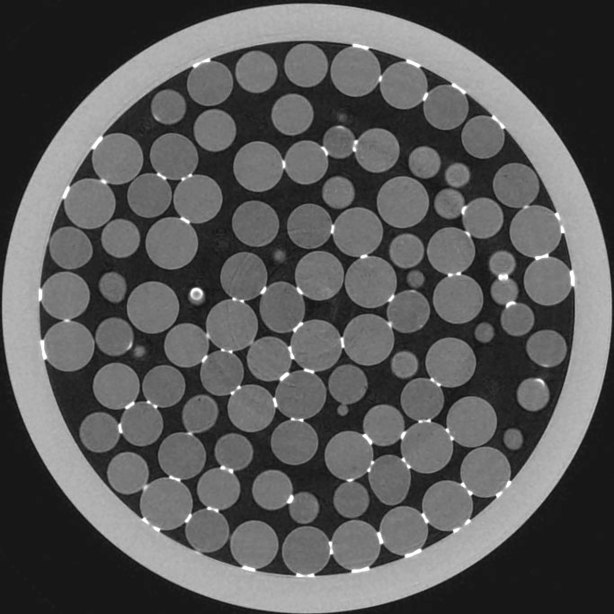
In this experiment you will learn the basics of the generation of X-rays and their interaction with matter and how these are used in computer-assisted X-ray tomography, a non-destructive three-dimensional imaging method. First, you will investigate the dependence of the intensity of the X-rays on the anode current, the acceleration voltage and the integration time of the scintillation detector. Using absorption images of wedges made of various materials, you will determine the attenuation of X- rays in matter and compare it with literature values. Using model samples, you will learn the basic concept of how three-dimensional images can be reconstructed from 2D projections. At the end of the experiment you will have the opportunity to record a complete tomography of an object of your choice.
The experiment is only suitable for master students. Supervision can be provided in German or English.
Martin Brinkmann
Building E 2.9, Room 3.04
Phone: +49 681 302-71722
martin.brinkmann(at)physik.uni-saarland.de
Biophysics

In our experiment, we work with an HL60 cell line that has been specifically differentiated into neutrophilic cells. We are investigating the impact of inhibiting Myosin II on their migration speed. To conduct this study, we utilize a PDMS microchannel system and employ fluorescence microscopy for experiment execution and result analysis. The experiment is divided into three parts: the first involves the fabrication of microchannels, the second part focuses on cell preparation, and the third part entails microscopic examination. The data analysis will be explained in a subsequent session. Consequently, the experiment provides insights into cell culture, microscopy, analysis, and general laboratory practices.
The experiment is suitable for all courses of study. Supervision can be provided in German or English.
Gagan Sharma
Building D 2.2, Room A2.12
Phone: +49 681 302-9 300 460
gash00001(at)uni-saarland.de
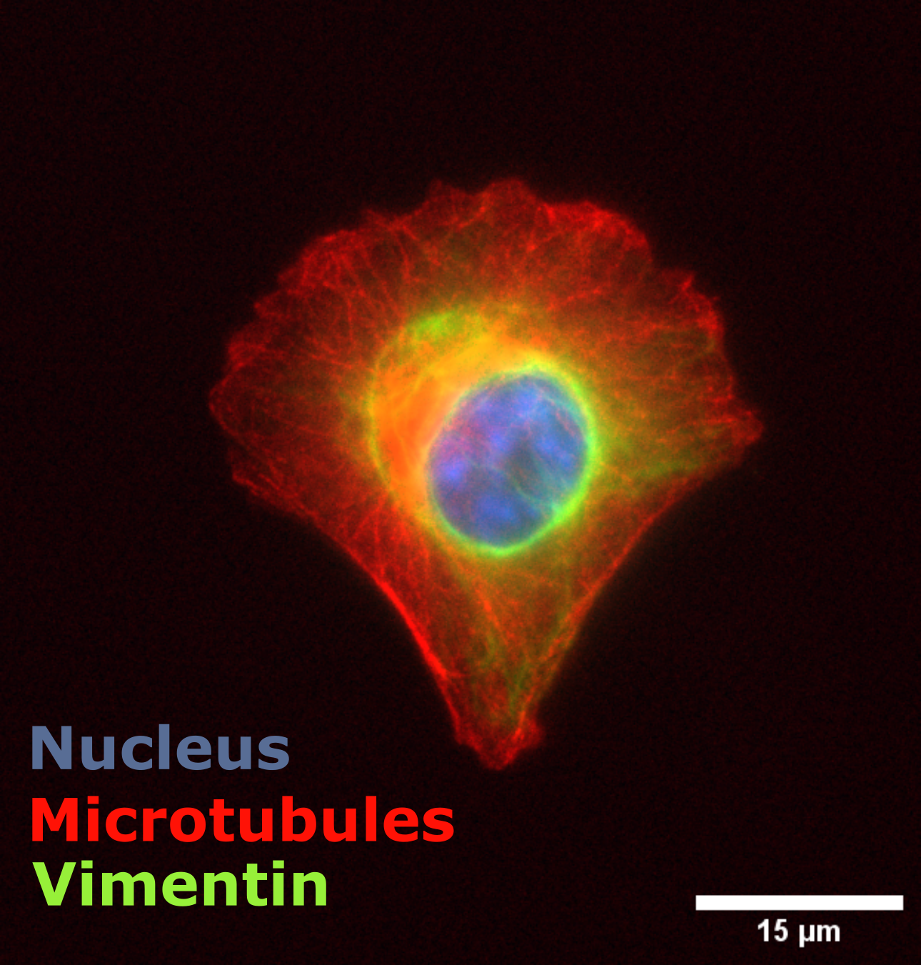
As the smallest building blocks of life, cells must be able to perform many different tasks to enable life as we know it. These include the movement of cells, the absorption of external substances, the processing of external signals and much more.
In order to be able to perform these tasks, they sometimes have to reorganize and realign their entire inner life. For example, nutrients that are absorbed by the intestinal wall should only be carried into the blood and not return to the intestine. In order for this to work, intestinal cells have an asymmetrical structure that allows them to determine a transport direction.
Such asymmetries can also be artificially enforced in cells. In the practical experiment "Cell Polarity", you will force cells into geometric shapes and at the same time investigate what effects this has on the filament network and the position of the organelles inside the cell.
The experiment is suitable for all courses of study. Supervision can be provided in German or English.
Carsten Baltes
Building D 2.2 (INM)
Phone: +49 681 302-9 300 460
s8cabalt(at)stud.uni-saarland.de
Dynamic Light Scattering (DLS) is a technique aiming to determine the size of particles using their diffusion coefficient. Experimentally, a laser beam enters a cuvette containing the analyzed sample, and the intensity of the scattered light is measured to determine the diffusion coefficient. In this experiment, polystyrene and silica beads of variable sizes will be used to investigate different regimes of the DLS.
The experiment is suitable for all courses of study. Supervision can be provided only in English.
Timothée Boutfol
Building B 2.1, Room 2.12
Phone: +49 681 302-68534
timothee.boutfol(at)uni-saarland.de
Microscopy methods

In this experiment you will learn the basics of atomic force microscopy (AFM) and carry out independent measurements on various samples. Surface topography and material properties can be investigated with resolutions in the nanometer range via the interactions between the measuring tip and the sample surface.
You will carry out AFM measurements on polymer samples and tooth enamel, among other things, and finally you will be able to scan your own samples.
The experiment is suitable for all courses of study. Supervision can be provided in German or English.
Jens Uwe Neurohr
Building E 2.9, Room 3.12
Phone: +49 681 302-71725
ju.neurohr@physik.uni-saarland

In this experiment, you will investigate the Brownian motion of beads in water. You will start with a simulation of the stochastic motion. Next you will observe the free Brownian motion of beads using light and dark field microscopy. The experiment will also show you typical imaging errors. Finally, you will look at Brownian motion in the harmonic potential of optical tweezers.
The experiment is suitable for all courses of study. Supervision can be provided in German or English.
Thomas John
Building E 2.6, Room 3.20
Phone: +49 681 302-2977
thomas.john(at)physik.uni-saarland.de
Solid state physics
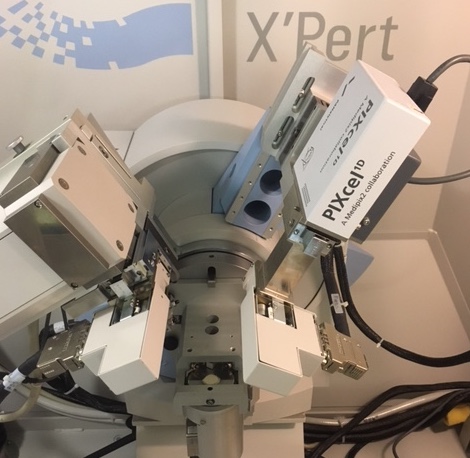
X-ray diffraction is a standard laboratory method for determining the structure of crystalline materials. The periodic arrangement of atoms in crystals leads to coherent scattering of photons (x-rays) that depends on the distance separating the atoms (lattice parameter),
on the size of the crystalline regions in which the atoms are located (grain size), and on fluctuations in atomic position about the expected position in the crystal lattice (strain).
In this experiment you will measure all three of these quantities for a nanocrystalline PdAu sample and nanocrystalline and a coarse-grained CeO2 sample.
The experiment is suitable for all courses of study. Supervision can be provided in German or English.
Anna Fuchs
Building E 2.6, Room 1.17
Phone: +49 681 302-2739
a.fuchs(at)physik.uni-saarland.de
Nuclear physics
This experiment offers an introduction into gamma radiation and spectroscopy of gamma ray energies. You will work with an ensemble of analog and digital signal processing. Furthermore you have the opportunity to get your hands on antimatter and exotic molecules and measure properties of nuclei. The established methods are for example used for identification of isotopes in radioactive samples and the characterization of defects or electromagnetic fields in crystal structures.
The experiment is suitable for all courses of study. Supervision can be provided in German or English.
Felix Maurer
Building E 2.6, Room 3.20
Phone: +49 681 302-2977
felixmilan.maurer(at)uni-saarland.de
Theoretical physics
The project 'Computed tomography' focusses on the mathematical basics for generating sinograms and reconstructing the original objects (Radon transform), which are then implemented and tested in Python or C.
The experiment is suitable for all courses of study. Knowledge of programming (Python, C or C++) and of linear algebra and numerics are required. Supervision can be provided in German or English.
Christian Hoffmann
Building E 2.6, Room 1.05
Phone: +49 681 302-4218
chhof(at)lusi.uni-sb.de
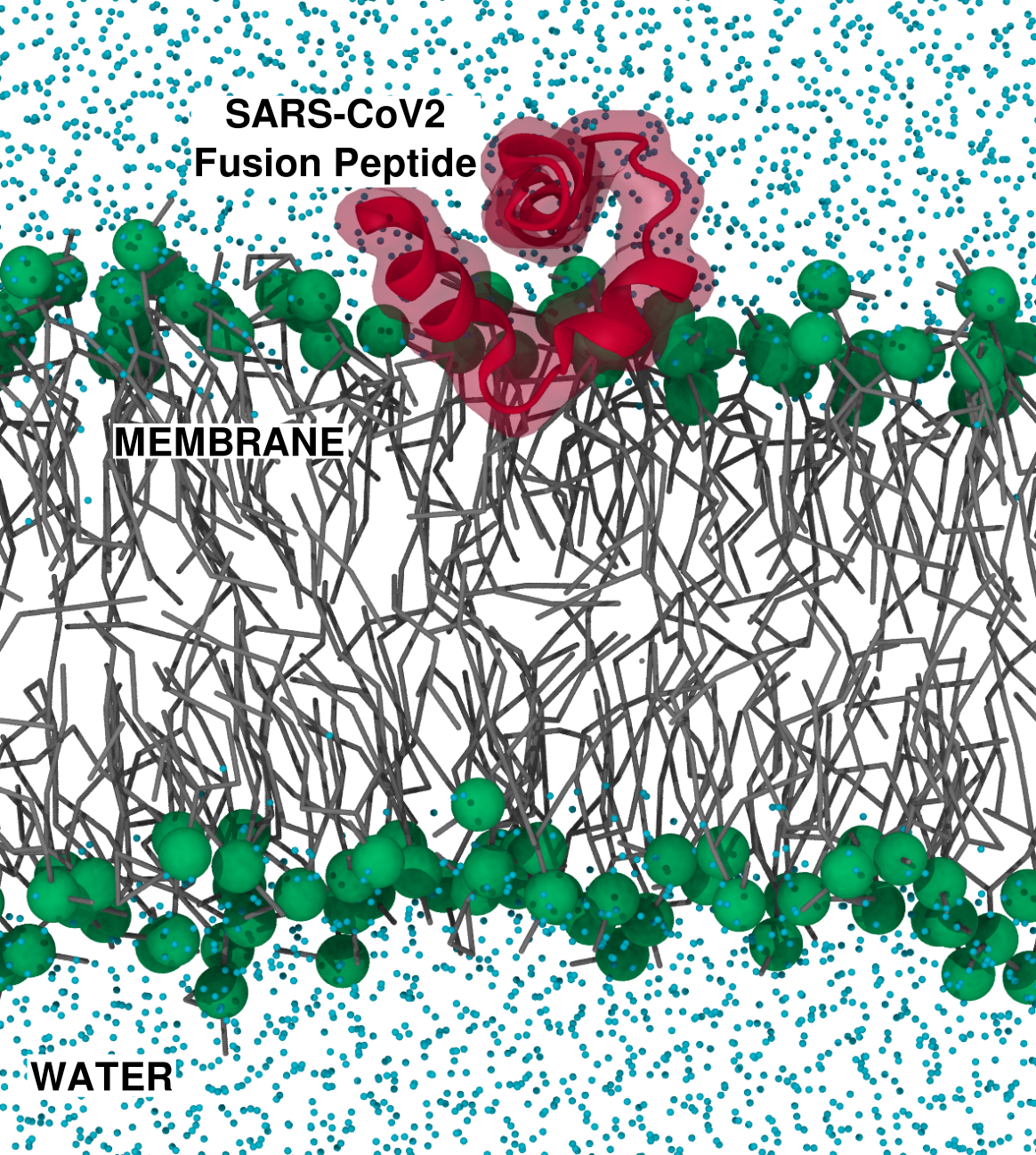
Integrating experiments and simulations strengthens multi- and cross-disciplinary approaches for solving complex biological problems. In biosciences, molecular dynamics simulations provide significant value to experimental research, particularly in super-resolution bioimaging, which has achieved spatial resolutions of approximately 20-50 nm. When exploring biological phenomena at scales beyond the reach of experimental techniques—such as the dynamics and interactions of individual biomolecules—molecular dynamics simulations and theoretical approaches are the most effective tools. In our practical class, you will apply both conventional and enhanced sampling molecular dynamics simulation techniques to investigate how the SARS-CoV-2 fusion peptide interacts with lipid membranes during viral infection.
The experiment is suitable for all courses of study. Supervision can be provided in English.
Alejandro Martínez León
Building E 2.6, Room 1.08
Phone: +49 681 302-3959
alejandro.martinezleon(at)uni-saarland.de